
This guide meticulously details dissection procedures for key domestic animals – horses, oxen, goats, and sheep – essential for veterinary curricula․
It’s crafted for both novice students and seasoned professionals seeking a robust anatomical understanding․
Purpose of the Dissection Guide
The primary aim of this dissection guide is to furnish a meticulously detailed, step-by-step resource for exploring the anatomy of the horse, ox, goat, and sheep․ Specifically designed for veterinary students navigating their professional curriculum, it also serves as a valuable reference for practicing veterinarians․
This edition, the third, intends to provide comprehensive anatomical knowledge through practical application․ It’s not merely about identifying structures, but understanding their relationships and spatial arrangements within these species․ The inclusion of full-color photographs enhances the learning experience, offering clear visual guidance throughout each dissection․
Ultimately, the guide strives to empower users with the skills and confidence needed to accurately interpret anatomical structures, crucial for successful diagnostic and surgical procedures in equine and ruminant medicine․ It bridges the gap between theoretical knowledge and practical expertise․
Target Audience: Veterinary Students & Professionals
This dissection guide is principally intended for first-year veterinary students embarking on their professional curriculum․ It’s designed to complement classroom learning by providing a hands-on, practical approach to anatomical study․ The detailed instructions and accompanying visuals are tailored to support those new to dissection techniques and anatomical terminology․
However, the scope extends beyond the student population․ Practicing veterinarians will also find this resource invaluable for refreshing their anatomical knowledge, preparing for complex surgical procedures, or investigating unusual anatomical variations․
The guide’s comprehensive nature and clear presentation make it accessible to individuals with varying levels of anatomical experience, serving as a reliable reference tool throughout their careers․ It aims to foster a deeper understanding of equine and ruminant anatomy for all users․
Scope of Species Covered: Horse, Ox, Goat, and Sheep

This guide provides comprehensive dissection coverage for four crucial domestic animal species: the horse, ox (cattle), goat, and sheep․ Recognizing the anatomical variations between these animals is paramount for veterinary practice․ The horse, representing the equine species, receives detailed attention due to its unique anatomical features and clinical significance․
Similarly, the ruminant digestive system – exemplified by the ox, goat, and sheep – is thoroughly explored, highlighting the complexities of their multi-compartment stomachs and associated structures․
By encompassing these four species, the guide offers a broad foundation in comparative veterinary anatomy, enabling students and professionals to confidently approach a wide range of clinical cases and anatomical investigations․ The detailed approach ensures a robust understanding of each species’ specific anatomy․

Essential Dissection Tools & Equipment
Successful dissection requires scalpels, scissors, forceps, retractors, and specialized pans․ These instruments, alongside detailed guides, facilitate precise anatomical exploration and understanding․
Scalpels and Blades
Scalpels are fundamental for initial skin incisions and deeper tissue separation during dissection․ A variety of blade shapes – from robust to delicate – are crucial for navigating diverse anatomical structures in equine and ruminant species․ The Guide to Dissection of the Horse and Ruminants emphasizes selecting appropriate blade types based on tissue density and desired precision․
Disposable scalpels with replaceable blades are recommended for maintaining sterility and sharpness throughout the process․ Regularly changing blades ensures clean cuts and minimizes tissue damage․ Different handle designs cater to varying hand sizes and dissection styles․ Careful handling and proper disposal of used blades are paramount for safety․ The guide likely details specific blade recommendations for different anatomical regions, optimizing dissection efficiency and accuracy․
Dissecting Scissors
Dissecting scissors are indispensable for precise tissue sectioning, particularly around nerves, vessels, and delicate structures encountered during equine and ruminant dissections․ The Guide to Dissection of the Horse and Ruminants likely highlights the importance of having a range of scissor types available․
Blunt-tipped scissors are ideal for separating muscle layers and protecting underlying tissues, while sharp-tipped scissors excel at cutting through tougher connective tissues․ Metzenbaum scissors, known for their fine blades and curvature, are particularly useful for intricate dissections․ Mayo scissors, with their heavier build, are suited for cutting denser tissues․ Maintaining scissor sharpness is vital for clean, controlled cuts, minimizing trauma and ensuring accurate anatomical identification․ Proper scissor care extends their lifespan and enhances dissection quality․
Forceps and Hemostats
Forceps and hemostats are crucial for grasping, manipulating, and temporarily clamping tissues and vessels during dissection of horses and ruminants․ The Guide to Dissection of the Horse and Ruminants emphasizes their role in controlled anatomical exploration․
Various types are essential: smooth forceps for delicate tissue handling, toothed forceps for firmer grip, and hemostats (like Kelly or Crile) for clamping blood vessels to control bleeding․ Different sizes and curvatures cater to varying anatomical locations․ Proper use prevents tissue damage․ Hemostats aren’t for prolonged clamping, as they can cause ischemia․ Understanding the appropriate application of each instrument, as detailed in the guide, ensures efficient and accurate dissection, revealing intricate anatomical relationships within the animal․
Retractors
Retractors are indispensable for maintaining clear visibility during dissection, particularly within the expansive cavities of equine and ruminant bodies․ The Guide to Dissection of the Horse and Ruminants highlights their importance in exposing deeper structures without causing undue tissue trauma․
Various designs – handheld, self-retaining (like Senn or Weitlaner), and specialized – are utilized․ Handheld retractors require continuous manual pressure, while self-retaining types lock into position, freeing hands for dissection․ Choosing the correct retractor size and shape is vital for effective exposure․ Careful application prevents damage to underlying tissues and organs․ The guide stresses the importance of gentle, controlled retraction to reveal anatomical details accurately, facilitating a comprehensive understanding of the animal’s internal architecture․
Dissection Pans and Instruments
Essential for organized dissection, robust dissection pans provide a contained workspace, preventing fluid spillage and maintaining a clean environment․ The Guide to Dissection of the Horse and Ruminants emphasizes the need for appropriately sized pans to accommodate large specimens․
Beyond pans, a suite of supplementary instruments is crucial․ These include probes for delicate tissue separation, pins for securing skin flaps, and measuring tools for accurate anatomical documentation․ The guide likely details proper instrument handling techniques to maximize efficiency and minimize tissue damage․ Maintaining sharp blades and clean instruments is paramount for precise dissections․ Proper organization of these tools streamlines the process, allowing students and professionals to focus on anatomical exploration and learning․
Regional Anatomy: Equine Dissection
This section systematically guides users through equine anatomy, covering the head, limbs, and abdominal/pelvic cavities with detailed instructions and illustrative imagery․
Equine Head and Neck Dissection
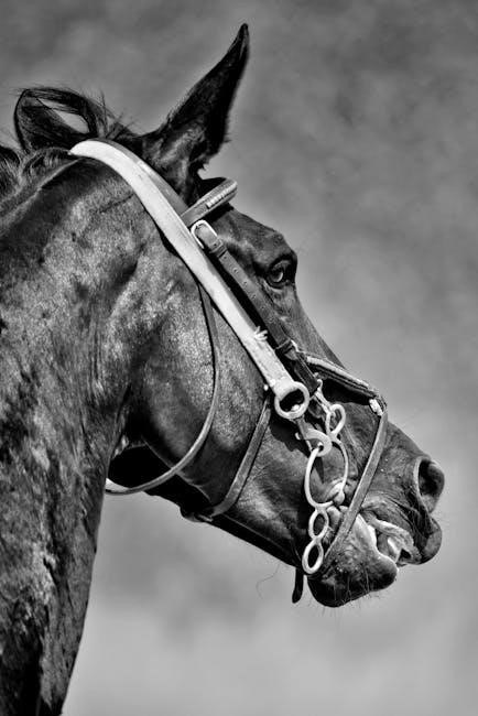
The equine head and neck dissection, as detailed in Abbott’s guide, begins with a careful examination of the superficial muscles responsible for facial expression and mastication․ Step-by-step instructions facilitate the identification of key structures like the masseter, temporalis, and various cutaneous muscles․
Full-color photographs aid in visualizing the intricate network of nerves and blood vessels supplying the region․ Dissection progresses to reveal the hyoid apparatus, salivary glands, and the nasal passages, emphasizing the unique anatomical features of the equine head․ The guide provides clear guidance on safely exposing the cranial nerves and tracing their pathways․
Students will learn to identify the major arteries and veins, including the external and internal carotid arteries, and the jugular vein․ Attention is given to the anatomy of the larynx and pharynx, crucial for understanding equine respiratory function․ This section prepares students for more complex dissections by building a solid foundation in equine head and neck anatomy․
Equine Thoracic Limb Dissection
The dissection of the equine thoracic limb, guided by Abbott’s detailed illustrations, commences with the superficial muscles – brachiocephalicus, trapezius, and others – responsible for shoulder and upper arm movement․ The guide emphasizes careful skinning and muscle reflection to preserve underlying structures․
Students will systematically explore the muscles of the forearm and carpus, identifying tendons and ligaments crucial for locomotion․ Full-color photos clearly demonstrate the location of major arteries, veins, and nerves, including the brachial artery and median nerve․ Attention is directed towards the suspensory apparatus, a unique feature of the equine limb․
The dissection extends to the foot, revealing the digital flexor tendons and navicular bone․ Step-by-step instructions ensure accurate identification of all anatomical components․ This section provides a comprehensive understanding of the biomechanics and common injuries affecting the equine thoracic limb․
Equine Abdominal Cavity Dissection
Dissection of the equine abdominal cavity, as detailed in the 3rd edition, begins with a careful midline incision, exposing the peritoneal space․ The guide stresses the importance of identifying the layers of the abdominal wall and preserving key vessels during initial opening․
Students will systematically explore the digestive tract – stomach, small and large intestines – noting their anatomical relationships and mesenteries․ Detailed photographs illustrate the liver, spleen, pancreas, and kidneys, highlighting their lobar structure and vascular supply․ The urinary bladder and associated structures are also carefully examined․
Emphasis is placed on identifying the major abdominal arteries and veins, alongside the innervation of the gut․ Step-by-step instructions facilitate the tracing of the digestive pathway and understanding of visceral function within the equine abdomen․
Equine Pelvic Limb Dissection
The 3rd edition’s guide to equine pelvic limb dissection begins with careful skinning and muscle layer removal, revealing the underlying skeletal structure; Detailed illustrations aid in identifying the gluteal muscles, hamstrings, and the quadriceps femoris group, emphasizing their origins and insertions․
Students will meticulously dissect the stifle joint, examining the ligaments and menisci․ The guide provides clear instructions for tracing the major nerves and blood vessels supplying the limb, including the femoral artery and sciatic nerve․ Attention is given to the digital flexor tendons and associated sheaths․
Further dissection focuses on the hock joint, foot, and associated ligaments․ Step-by-step photos demonstrate proper technique for exposing the navicular bone and surrounding structures, crucial for understanding lameness issues in the horse․
Regional Anatomy: Ruminant Dissection
This section expertly guides users through ruminant anatomy – ox, goat, and sheep – with detailed dissections․ It’s a crucial resource for comparative anatomical study and practical skill development․
Ruminant Head and Neck Dissection
Dissecting the ruminant head and neck requires a systematic approach, beginning with superficial muscle identification․ Abbott’s guide provides step-by-step instructions and full-color photographs illustrating key structures like the masseter, temporalis, and sternohyoid muscles․ Careful attention should be paid to the hyoid apparatus and its associated ligaments, crucial for understanding ruminant deglutition․
The guide details the intricate network of nerves and blood vessels supplying the head, including the facial, vagus, and glossopharyngeal nerves․ Students will learn to identify the major salivary glands – parotid, mandibular, and sublingual – and trace their ducts․ Furthermore, the dissection extends to the nasal cavity, oral cavity, and pharynx, emphasizing the unique anatomical adaptations of ruminants for their specialized digestive processes․ Precise identification of bony landmarks is also highlighted․
Ruminant Thoracic Limb Dissection
Dissection of the ruminant thoracic limb, as detailed in Abbott’s guide, begins with superficial muscle groups – brachiceps, triceps brachii, and biceps brachii․ The guide’s illustrations clearly demonstrate the origins and insertions of these muscles, alongside the underlying brachialis․ Careful attention is given to the shoulder joint and its supporting ligaments, vital for understanding limb movement․
Students will then progress to dissecting the forearm, identifying muscles like the flexor and extensor carpi radialis․ The guide emphasizes the importance of tracing nerves and vessels – the brachial artery and median nerve – throughout the limb․ Detailed instructions cover the carpus, metacarpus, and digits, highlighting the unique features of ruminant hooves․ Finally, the guide aids in understanding suspensory apparatus and digital flexor tendons․
Ruminant Abdominal Cavity Dissection (Ox)
Dissecting the bovine abdominal cavity, as guided by Abbott’s text, necessitates a systematic approach․ Begin by exposing the rumen, reticulum, omasum, and abomasum – the four compartments defining ruminant digestion․ The guide provides detailed illustrations of their anatomical relationships and vascular supply․
Students will then identify the liver, pancreas, and spleen, noting their positions relative to the digestive tract․ The dissection continues with the intestines, differentiating the small and large intestines, and tracing their mesenteries․ Crucially, the guide highlights the unique features of the bovine kidney and adrenal glands․ Finally, the text aids in locating major vessels like the aorta and vena cava, alongside associated lymph nodes․

Ruminant Abdominal Cavity Dissection (Goat/Sheep)
Dissecting the abdominal cavity of goats and sheep, as detailed in Abbott’s guide, reveals similarities and differences compared to the bovine anatomy․ Begin by carefully exposing the four stomach compartments – rumen, reticulum, omasum, and abomasum – noting their relative sizes and positions․ The text emphasizes identifying the greater and lesser omentum․
Students will then locate the liver, spleen, and pancreas, observing their vascular connections․ The dissection proceeds to the intestines, differentiating the small and large intestines, and identifying the cecum․ Importantly, the guide illustrates the kidney and adrenal gland locations․ Finally, tracing the aorta, vena cava, and associated lymph nodes completes the dissection, aided by full-color photos within the guide․
Ruminant Pelvic Limb Dissection
The ruminant pelvic limb dissection, as guided by Abbott’s 3rd edition, begins with superficial muscle exposure․ Students carefully remove skin and fascia to reveal the gluteal muscles, hamstrings, and gastrocnemius․ Detailed, step-by-step instructions aid in identifying the sciatic nerve and associated vasculature․
Further dissection focuses on the stifle joint, revealing the cruciate ligaments and menisci․ The guide then directs attention to the hock joint, showcasing its complex structure․ Particular emphasis is placed on tracing the digital flexor tendons to the hoof․ Throughout, the text highlights anatomical variations common in goats and sheep․ Full-color photos provide crucial visual references, ensuring accurate identification of structures during the dissection process․
Specific Anatomical Structures & Dissection Techniques
This edition expertly guides users through musculoskeletal, visceral, nervous, and vascular systems, utilizing detailed instructions and full-color imagery for precise anatomical exploration․
Musculoskeletal System Dissection
Dissecting the musculoskeletal system within the horse and ruminants demands a systematic approach, beginning with superficial muscle identification and progressing to deeper structures․ Abbott’s guide provides step-by-step instructions, emphasizing accurate tissue separation to reveal muscle attachments, tendon pathways, and ligamentous connections․
Particular attention is given to the unique features of equine limb anatomy, including the splint bones and suspensory apparatus․ For ruminants, the guide highlights variations in muscle mass and skeletal proportions compared to the horse․
Full-color photographs illustrate key dissection landmarks, aiding in the identification of individual muscles, joints, and bony structures․ The text details techniques for safely removing muscle layers, exposing underlying nerves and blood vessels, and preserving anatomical relationships throughout the dissection process․ Understanding these structures is crucial for veterinary professionals․
Visceral Organs Dissection
Dissection of visceral organs in the horse and ruminants, as detailed in Abbott’s guide, requires careful technique to preserve anatomical relationships and identify key structures․ The guide emphasizes a systematic approach, beginning with the abdominal cavity and progressing through the thoracic cavity․
Special attention is given to the ruminant digestive system, highlighting the multi-compartment stomach and its associated vasculature and innervation․ Equine visceral anatomy is presented with detailed illustrations of the liver, kidneys, and intestines․
Step-by-step instructions and full-color photos facilitate the identification of organs, their attachments, and surrounding tissues․ The guide also covers techniques for tracing major blood vessels and nerves, providing a comprehensive understanding of visceral anatomy essential for veterinary diagnosis and surgery․
Nervous System Dissection
Abbott’s Guide to Dissection provides detailed protocols for exploring the nervous systems of both equine and ruminant species․ The guide stresses a methodical approach, starting with the brain and spinal cord, then progressing to peripheral nerves․ Careful dissection techniques are crucial for preserving delicate neural tissues․
Full-color photographs illustrate key landmarks and anatomical relationships, aiding in the identification of cranial nerves, the brachial plexus, and major spinal nerve roots․ The ruminant nervous system is compared and contrasted with that of the horse, highlighting species-specific differences․
Step-by-step instructions guide students through the process of tracing nerve pathways and identifying associated structures, fostering a deeper understanding of neurological function and potential clinical implications․
Vascular System Dissection
The Guide to Dissection offers comprehensive instructions for dissecting the arterial and venous systems of the horse, ox, goat, and sheep․ Emphasis is placed on identifying major vessels – aorta, vena cava, pulmonary arteries and veins – and their branching patterns․
Detailed illustrations showcase the location of key vascular structures within the thoracic and abdominal cavities, as well as the limbs․ Students learn to trace the course of vessels and identify associated lymphatic drainage pathways․
Step-by-step guidance is provided for careful dissection, ensuring preservation of vessel integrity․ The guide highlights species-specific variations in vascular anatomy, crucial for understanding clinical conditions and surgical approaches․